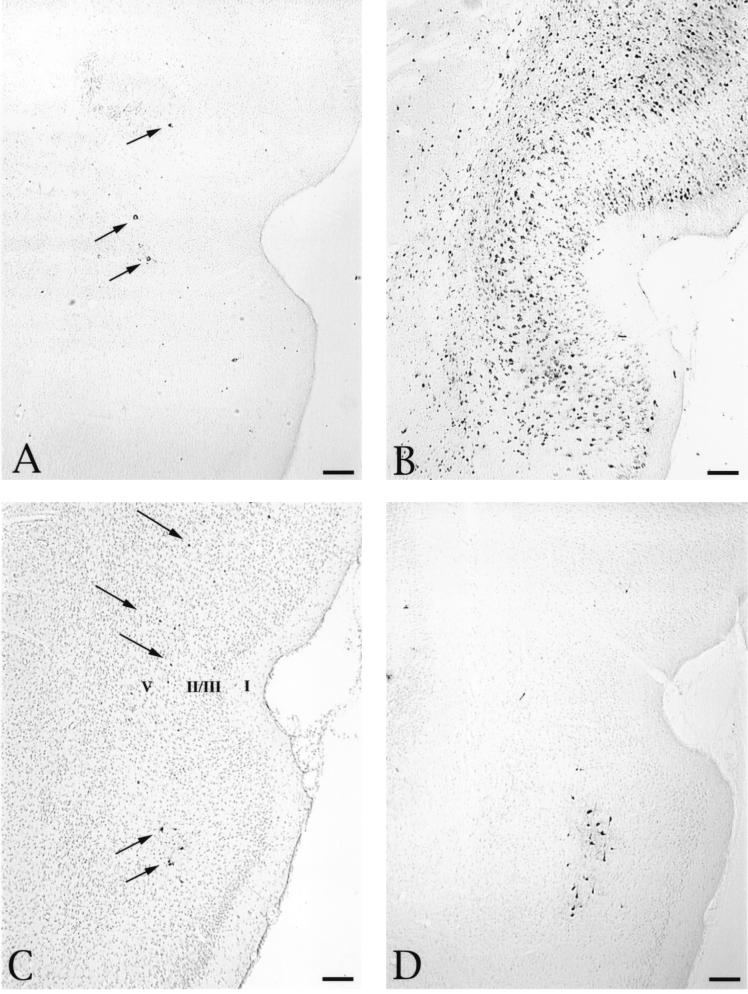
FIG. 4.
Distribution of infected neurons in the perirhinal cortex following injection of PRV-Becker or PRV-Bartha into the PFC. The distribution of infected neurons approximately 27 (A) and 48 (B) h after injection of 200 nl of PRV-Becker into the PFC is shown. At the shorter survival time, infected neurons are few and confined to lamina V of the perirhinal cortex (arrows). A substantially larger group of infected neurons are present in the perirhinal cortex 47 h after injection and extend through all cortical laminae. Scattered infection of layer V neurons is also observed 48 h after injection of 100 nl of either PRV-Becker (C) or PRV-Bartha (D) (arrows). The section illustrated in panel C was counterstained with cresyl violet to aid in the identification of cortical laminae, which are numbered according to the criteria defined by Swanson (43) to serve as points of orientation for defining the laminar disposition of infected neurons in the sections that were not counterstained. Bars, 200 μm.

