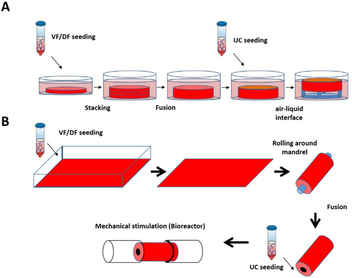
Figure 4.
Schematic protocol of urethral substitute production by the self-assembly method (flat and tubular models). (A) Preparation of flat tissue (patch): The stromal cells are seeded on 6-well plates containing a paper anchor used to limit contraction and to facilitate tissue manipulation. A support stabilizes this anchoring. The cells are cultured in a medium supplemented with 50 μg/mL of ascorbate. On day 28, the sheets are detached from the plastic, and three sheets are superimposed to form a thicker tissue subjected to limited compression to allow the fusion of the cell sheets in 4 days. Then, the urothelial cells are seeded on the upper part of the construct and cultivated for seven days in submerged condition. The constructions are then raised at the air/liquid interface using a support for an additional 21 days to allow the epithelial cells to mature. (B) Diagram of the “tubular” model production by the self-assembly technique. Classic tubular self-assembly technique. The dermal fibroblasts are seeded in a cell culture plate covered with gelatin. They are grown for 28 days in the presence of ascorbate. The stromal sheet formed is tightly wound around a cylindrical mandrel. The resulting construction is cultivated for four days to ensure the fusion of the sheets. Once the fusion is complete, the mandrel can be removed, and the tubular structure can be perfused to be seeded with urothelial cells. The tube is matured in a bioreactor.

