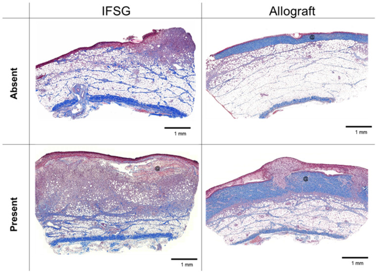
Figure 3.
Mason’s trichrome histology at day 7 with granulation tissue formation. Representative images of biopsy punches harvested at the day-7 time point prior to skin graft application that align with the pathologist scoring of granulation tissue shown in Table 2. Black scale bars = 1 mm. @ = residual pieces of treatment in wound bed. IFSG = intact fish skin graft.

