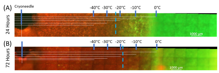
Figure 3.
Fluorescent micrographs of the PDAC-TEMs following single 5 min freeze. At 24 h (A) and 72 h (B) post-freeze, the extent of PANC-1 cell death in TEMs was evaluated via fluorescence microscopy following staining with Calcein-AM (green, live) and propidium iodide (red, dead). Using the Zeiss ZEN software, measurements were made, and then temperatures from the corresponding thermocouples were overlayed. Analysis revealed complete PANC-1 cell destruction was attained following exposure to a temperature of ~−20 °C or colder.

