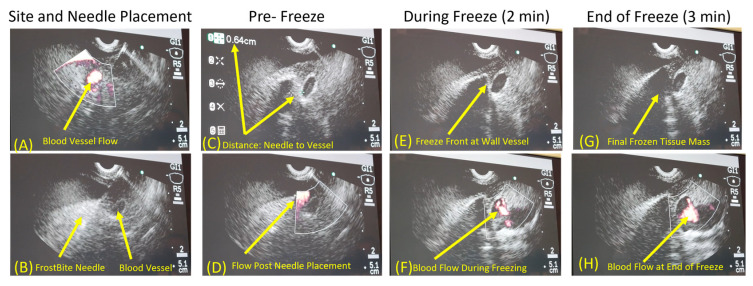
Figure 5.
Endoscopic ultrasound Doppler imaging of a single 3-minute freeze protocol next to a blood vessel. Images demonstrate blood flow through the vessel prior to FrostBite placement (A), visualization of FrostBite needle placement (B), measurement of the distance between the blood vessel and FrostBite (C), and Doppler blood flow prior to freeze initiation (D). Lesion visualization of iceball approaching the blood vessel (E) and blood flow in vessel (F) following 2 min of freezing. Iceball growing around blood vessel (G) and Doppler assessment of blood flow (white region) in vessel (H) at the end of the 3 min freeze protocol. Images show that when FrostBite was placed ~0.6 cm from a blood vessel and a 3 min freeze protocol was performed, the frozen tissue mass sculpted around the blood vessel, and the freezing process did not impact blood flow through the vessel throughout the freeze procedure.

