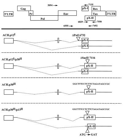
FIG. 1.
Mutations constructed in ACH resulting in the loss of expression of p12I, p30II, and p13II. At the top is a schematic drawing of the ORFs in the HTLV-1 genome and the LTRs. The four figures below this represent the transcripts encoding each potential protein product. The triangles indicate the positions of the mutations, and the dotted lines represent the regions of the genome which are removed by splicing. The nucleotide positions are numbered as for the parental ACH clone. The primers used for the confirmation of the mutations (Fig. 2 and 3) are indicated and are designated by the position of the 5′ end of the primer.

