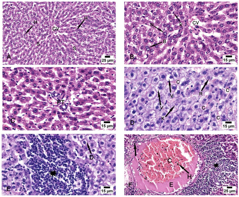
Figure 3.
Photomicrographs of the liver tissue. (A–C) Group I shows the normal architecture of the liver tissue around the central vein (CV) and the portal triad (PT). The central vein is surrounded by radiated cords of hepatocytes (H) and hepatic sinusoids (S). The Kupffer cells (K) are seen attached to the endothelium of the sinusoids. The nuclei appeared vesicular with prominent nucleoli (N), and some binucleated cells are noted (B). (D–F) Group II shows degeneration of the hepatocytes with loss of their normal laminar pattern. Necrosis in the form of nuclear karyolysis (K) and karyorrhexis (R) with sinusoidal congestion (C) and vacuolar degeneration of the hepatocytes (V) are seen. Inflammatory cellular infiltration (asterisk), dissolution of the cytoplasm (D), dilated and congested (C) portal vein with portal edema (E), periportal fibroplasia (arrow), and hemosiderosis (H) are observed. (H & E; (A,F) ×200; (B–E) ×400).

