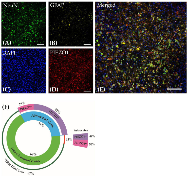
Figure 2.
Cortical cultures derived from embryonic mice on DIV21 stained for (A) Neuronal Nuclear Protein (NeuN), (B) Glial Fibrillary Acidic Protein (GFAP), (C) DAPI, and (D) PIEZO1. (E) Merged image of all channels. Images were taken at 40× magnification. Scale bar = 50 µm. (F) The pie chart reflects the percentage distribution of the cell types in the cortical culture and their co-localization percentage with PIEZO1.

