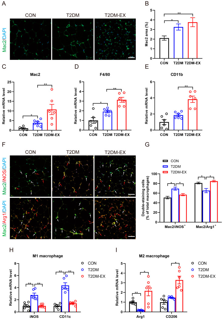
Figure 5.
The effects of exercise intervention on macrophage infiltration and polarization in T2DM mice. (A) Representative images of Mac2 immunofluorescence staining of iWAT in control, T2DM, and T2DM-EX mice, scale bar = 50 μm. (B) Percentage of Mac2-positive area in total image, 3 samples per group, 6 fields per section. (C–E) The mRNA expression levels of pan-macrophage markers (Mac2, F4/80, and CD11b). (F) Representative images showing the double-positive staining of Mac2 and iNOS, Mac2 and Arg1. Scale bar = 50 μm. White arrows indicate Mac2 single-positive cells; red arrows indicate the double-positive cells. Green: Mac2; red: iNOS or Arg1; blue: nucleus stained by DAPI. (G) The ratio of double-positive cells to Mac2 single-positive cells. M1 represents Mac2+iNOS+ and M2 represents Mac2+Arg1+. Three samples per group, 6 fields per section. (H) The mRNA levels of M1-type macrophage markers (iNOS, CD11c) in iWAT. (I) The mRNA levels of M2-type macrophage markers (CD206, Arg1) in iWAT. * p < 0.05, ** p < 0.01. Data are expressed as mean ± SEM.

