Figure 3.
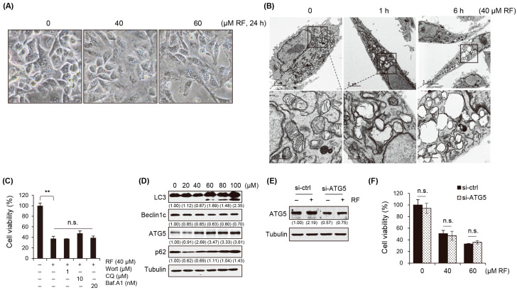
RF-induced autophagy was unrelated to cell death. (A) Morphological alterations in A549 cells observed under a microscope after exposure to 40 µM RF. (B) Transmission electron microscopy images of A549 cells treated with 40 µM RF for 1 or 6 h. (C) Cell viability of A549 cells pre-treated with the autophagy inhibitors wortmannin (Wort), chloroquine (CQ), and bafilomycin A1 (Baf.A1) were pre-treated for 1 h and then treated with 40 μM RF for 24 h using the MTT assay. (D) Western blot for autophagy-related genes in A549 cells treated with RF at the indicated concentrations for 24 h. (E) Western blot for ATG5 in non-targeted siRNA or ATG5 siRNA-mediated gene knockdown A549 cells in presence or absence of RF. (F) Cell viability of mock and ATG5 knockdown cells treated with RF at the indicated concentrations for 24 h using MTT. All data are expressed as the mean ± SD obtained from at least three independent experiments. Statistical differences were assessed using one-way ANOVA. ** p < 0.01 compared to the control. RF, rotundifuran; n.s., not significant.

