Figure 5.
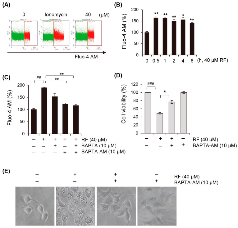
RF increased cytosolic calcium concentration. (A) Flow cytometry analysis of A549 cells treated with 20 and 40 µM RF for 30 min and stained with 2 μg/mL Fluo-4 AM. (B) Changes in cytoplasmic calcium levels in A549 cells exposed to 40 μM RF at the indicated times, assessed through staining with Fluo-4 AM. (C–E) A549 cells were pre-treated with 10 µM BAPTA, 10 µM BAPTA-AM, or 10 µM BAPTA plus 10 µM BAPTA-AM after exposure to 40 µM RF. (C) Changes in intracellular calcium concentration analyzed with Fluo-4 AM. Cell viability evaluated using MTT. (E) Vacuole formation observed using an optical microscope. Statistical differences were assessed using one-way ANOVA. * p < 0.05 and ** p < 0.01 compared to the RF-treated group. ## p < 0.01 and ### p < 0.001 compared to the control. RF, rotundifuran.

