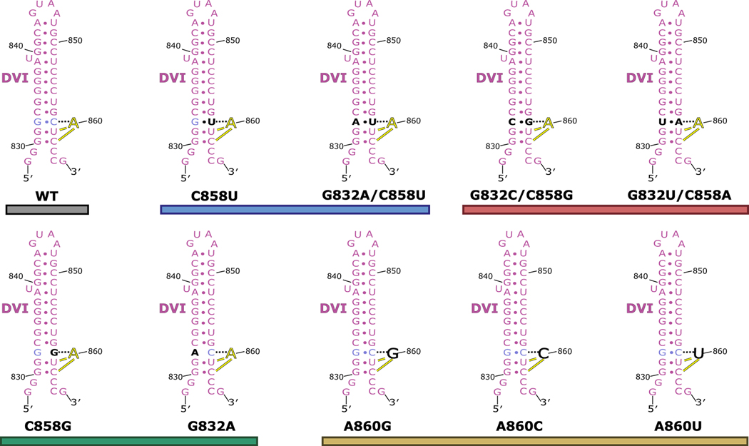
Extended Data Fig. 6. T.el4h mutant constructs used during in vitro splicing assay.
The secondary structure of DVI is shown for WT and the mutants tested in the in vitro splicing assay. The G832-C858 pair is shown in light blue and the branch-site adenosine (A860) is shown in yellow. Mutations are highlighted in black. The constructs are grouped and colored as seen in Figure 3B of the main text.

