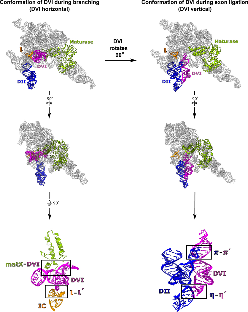
Figure 2. Conformational dynamics of the branch-site helix during RNA splicing.
DVI (magenta) is held in a horizontal position by the matX-DVI and ι-ι′ interactions during branching. These interactions stabilize the DVI helix in a conformation that brings the adenosine nucleophile to the 5′ SS. After branching, DVI swings 90° into a vertical position and is captured by the π-π′ and η-η′ interactions. Transition into this alternate conformation removes the newly formed lariat bond from the active site while simultaneously replacing it with the 3′ SS for exon ligation.

