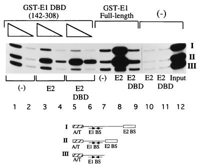
FIG. 3.
The E1 DBD interacts with the E2 DBD but not with the E2 activation domain. Three different probes containing a high-affinity E2 binding site distal to the E1 binding site (I), a high-affinity E2 binding site proximal to the E1 binding site (II), and only an E1 binding site (III) were mixed with either 0.5 (lane 1) or 0.25 (lane 2) ng of GST-E1(142–308) in 10-μl binding reactions; 2.0 ng of E2 (lanes 3 and 4) or 0.80 ng of the E2 DBD (lanes 5 and 6) was incubated with GST-E1(142–308) in the binding reaction. Probes bound by GST-E1 DBD were recovered by using glutathione-agarose beads. The recovered probes were analyzed on a 6% urea gel. Control reactions containing GST-E1 (full length) (6 ng) alone (lane 7) or together with full-length E2 (lane 8) or the E2 DBD (lane 9) were performed simultaneously.

