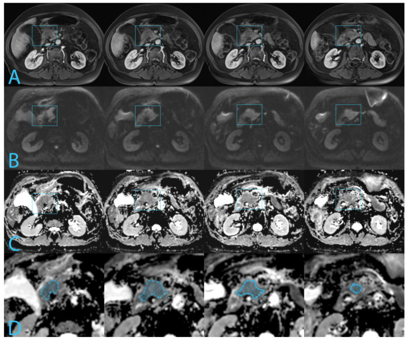
Figure 1.
Four slices of MR images depict a pT2N2 tumor measuring 35 mm in the pancreatic head, highlighted by the blue rectangle. The tumor exhibits an ADC p10 of 1038 µm2/s. Histopathologically, the tumor is classified as WHO moderately differentiated, Adsay G1 and a Kalimuthu tubulopapillary pattern. (A). T1-VIBE arterial phase. (B). DWI at b800 s/mm2. (C). ADC map. (D). Freehand regions of interest along the border of the tumor on the ADC map.

