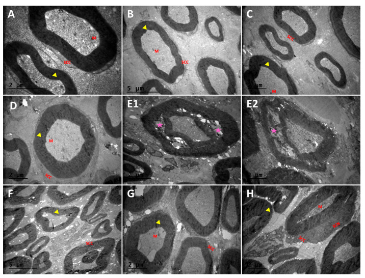
Figure 4.
Transmission electron micrographs (TEMs) of rat sciatic nerves. Myelinated nerve fibers (yellow arrowhead) and separation in the concentric lamellae of the myelin sheath and damaged axons (purple color star), SCC; Schwann cells, SCN; Schwann cell nucleus, M; mitochondria. (A); Control group, (B); propolis (P) group (C); quercetin (Q) group, (D); P + Q group, (E1,E2); diabetes mellitus (DM) group, (F); DM + P group, (G); DM + Q group, and (H); DM + P+ Q group. (Magnification, (A); ×20,000, (B,F); ×6000, (C–E1,G,H); ×10,000, and (E2); ×12,000).

