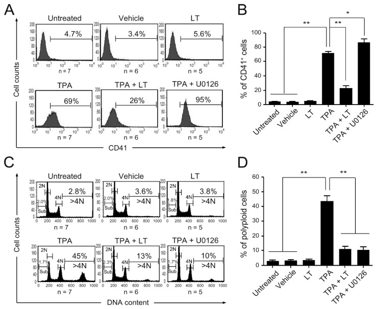
Figure 1.
Effect of LT and U0126 on megakaryocytic differentiation. HEL cells were untreated or treated with LT, TPA (10−8 M), TPA combined with LT, and TPA combined with U0126 (10 μM) for three days. HEL cells treated with 0.01% DMSO served as the vehicle control. The megakaryocytic-specific surface marker (CD41) expression cell percentages were monitored (A) and analyzed (B) using flow cytometry. Cells were stained with PI, and their DNA contents were characterized (C) and calculated (D) using flow cytometry. The numbers on the top right of (A,C) represent the percentages of CD41+ cells and polyploidy (DNA content > 4 N) cells in each group, respectively. The numbers on the left side of (C) represent the percentage of SubG1 cells in each group. Data are reported as mean ± the standard error of the mean (SEM). * p value < 0.05, ** p value < 0.01, compared with the indicated groups.

