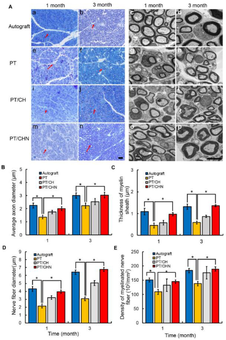
Figure 5.
(A) Cross-sections of regenerated nerves taken from nerve conduits implanted in rats after 1 and 3 months. Red arrows show nerve fibers. (a–d): Autograft group; (e–h): Poly(D, L-lactic acid)/β-tricalcium phosphate nerve conduits group; (i–l): Poly(D, L-lactic acid)/β-tricalcium phosphate nerve conduits/hyaluronic acid-chitosan group; (m–p): Poly(D, L-lactic acid)/β-tricalcium phosphate nerve conduits/hyaluronic acid-chitosan/ nerve growth factor group. (B) Axon diameters of regenerated myelinated nerve fibers. (C) Thicknesses of regenerated myelinated sheaths. (D) Diameters of regenerated nerve fibers. (E) Densities of regenerated myelinated nerve fibers. Data was analyzed by one-way ANOVA where p < 0.05 *. Reprinted from Xu et al. [109], copyright 2022, with permission of Elsevier.

