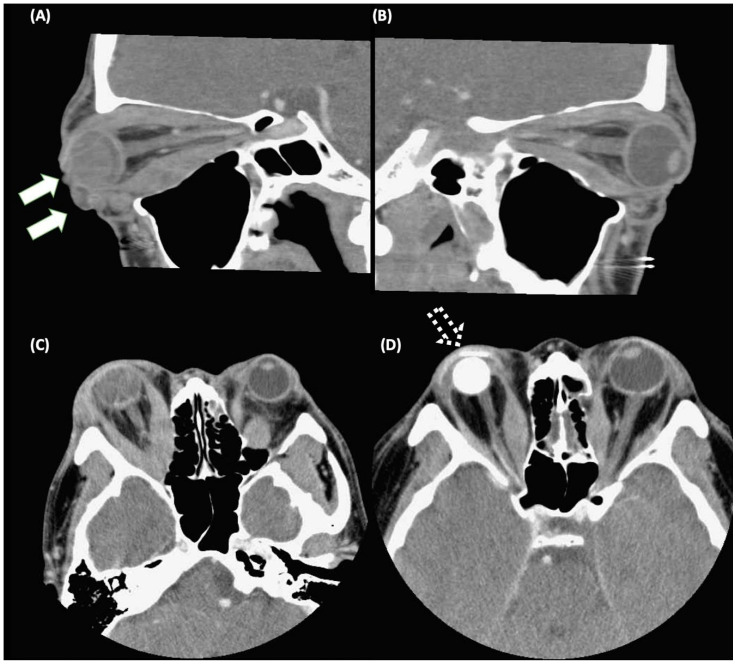
Figure 1.
Computed tomography images in a patient with Graves’ ophthalmopathy (GO). (A) Exposure keratopathy with eyeball rupture (solid arrows) due to severe extraocular muscle hypertrophy and adipogenesis in the right eye. (B) Extraocular muscle hypertrophy in the left eye. (C,D) The patient received an evisceration with an implant (as dashed arrows) of the right eye, and orbital decompression of the left eye. The pre-operative image and post-operative image were demonstrated in (C) and (D), respectively.

