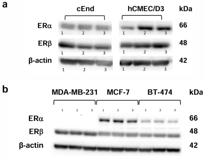
Figure 1.
Western blot analysis showing the protein expression patterns of the estrogen receptors (ERs) ERα (66 kDa) and ERβ (48 kDa). (a) The murine brain endothelial cell lines cEND (left) and the human brain endothelial cell line hCMEC/D3 (right) show the presence of both ERs in three experimental runs (1, 2, 3). (b) The breast cancer (BC) cell lines showed the presence of ERβ in MCF-7, BT-474 and MDA-MB-231 cells, whereas ERα was only detected in MCF-7 and BT-474. Again, three experimental runs were performed (1, 2, 3). β-actin (42 kDa) was used as a loading control.

