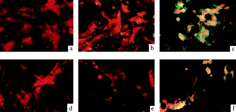
FIG. 2.
Immunofluorescence staining of IBDV-infected cells used to detect NS protein expression. CEF cells were infected with passage 1 (b) or passage 10 (e) rD78NSΔ mutant virus stock or with passage 1 (c) or passage 10 (f) rD78 virus stock at an MOI of 1. Uninfected CEF cells were used as negative controls (a and d). After 24 h postinfection, the cells were fixed and analyzed by immunofluorescence staining with rabbit anti-NS protein serum. Magnifications, ×400.

