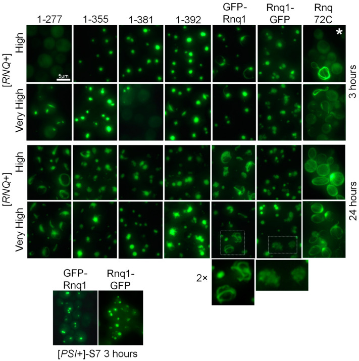
Figure 2.
Aggregation patterns of GFP-labeled Rnq1 protein constructs. [RNQ+] variants are indicated on the left, and Rnq1 constructs are shown on top. Two-fold magnification of the boxed areas is shown. The [PSI+]-S7 strain is ∆rnq1. *Ring-like aggregates in this sample were observed in only about 2% of cells.

