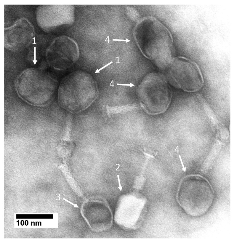
Figure 3.
Electron microscopy of MB-reacted, negatively stained phage T4. Phage T4 was incubated with 10.0 mM MB, as done in Figure 2, and then immediately negatively stained with 1.5% uranyl acetate [55]. Images were obtained with a JEOL100CX electron microscope (JEOL USA, Inc., Peabody, MA, USA) operated at 80 kV.

