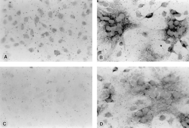
FIG. 2.
Detection of HPIV3 RNAs in the CSK framework. CV-1 cells, grown on coverslips, were infected with HPIV3 at 1 PFU/cell. The cells were treated with CSK buffer and hybridized with genome sense or antisense NP RNA labeled with digoxigenin. The coverslips were treated with anti-digoxigenin antibody coupled to alkaline phosphatase (Boehringer Mannheim), and the signal was detected with nitroblue tetrazolium and X-phosphate as the substrate. The coverslips were then mounted on slides and visualized under a phase-contrast microscope. Similarly treated mock-infected CV-1 cells served as the control. Mock-infected CV-1 cells (A) and HPIV3-infected cells (B) hybridized with antisense NP RNA (T7 transcript of NP cDNA clone in pGEM4Z) and mock infected CV-1 cells (C) and HPIV3-infected cells (D) hybridized with NP mRNA (SP6 transcript of NP clone in pGEM4Z) are shown.

