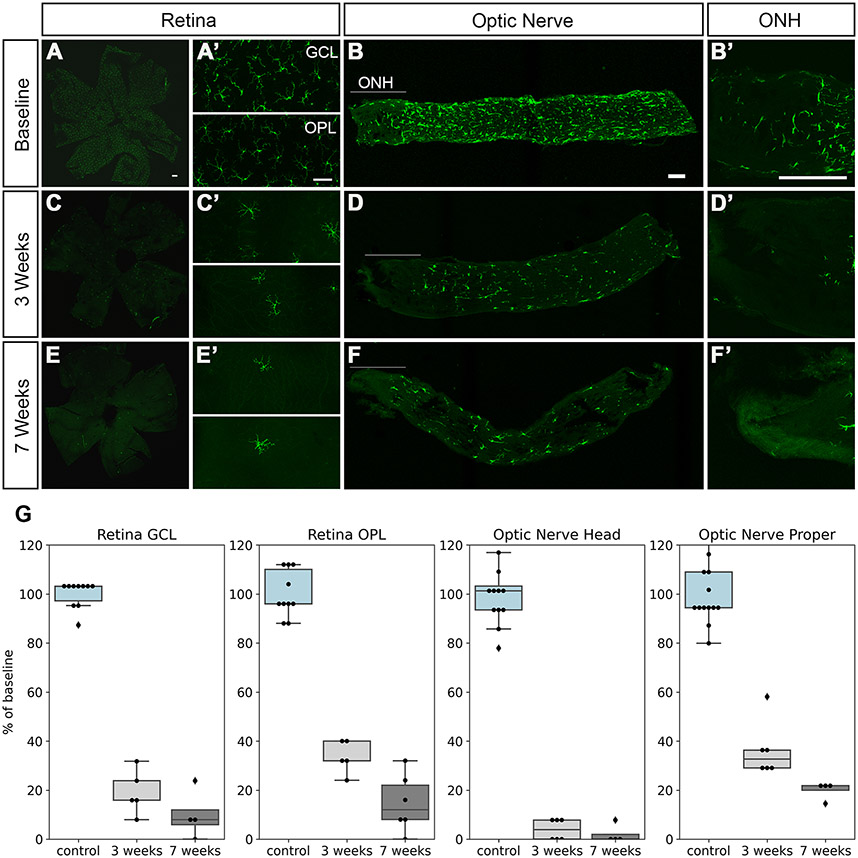
Figure 1.
Microglia depletion by PLX5622. Retinas, optic nerves, and optic nerve heads (ONH) from Cx3cr1-GFP mice were imaged at baseline, 3 weeks, and 7 weeks on the PLX5622 diet. At baseline, GFP+ cells with the morphology of resting microglia were present throughout the retina (A, A’), optic nerve (B), and optic nerve head (B’). The number of GFP+ cells decreased markedly at 3 weeks (C, D), and even more so at 7 weeks (E, F), but some labeled cells persisted, especially in the optic nerve proper (myelinated region of the optic nerve). Scale bars, 50 μm. G, quantification of GFP+ microglial cells. Baseline (control) shown in light blue, 3 weeks light gray, 7 weeks dark gray.

