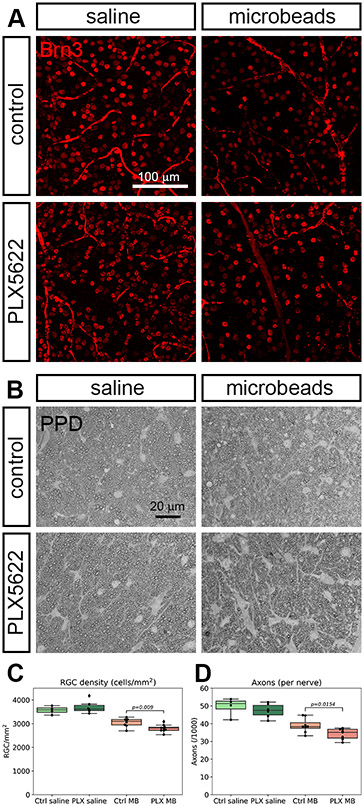
Figure 5.
Retinal ganglion cell and axon counts. A, retinal whole-mounts 4 weeks after injection with saline or microbeads were stained with the ganglion cell marker Brn3. Microbead injection leads to a loss of retinal ganglion cells that is more severe if the mice had received the microglia depletion diet. B, ganglion cell axons were stained with p-phenylenediamine (PPD) and counted. C, quantification of retinal ganglion cell density in the four experimental groups (regular diet and saline injection, light green; PLX5622 diet and saline injection, dark green; regular diet and microbead injection, light red; PLX5622 diet and microbead injection, dark red). D, quantification of the axon counts. Color code as in C.

