Figure 6.
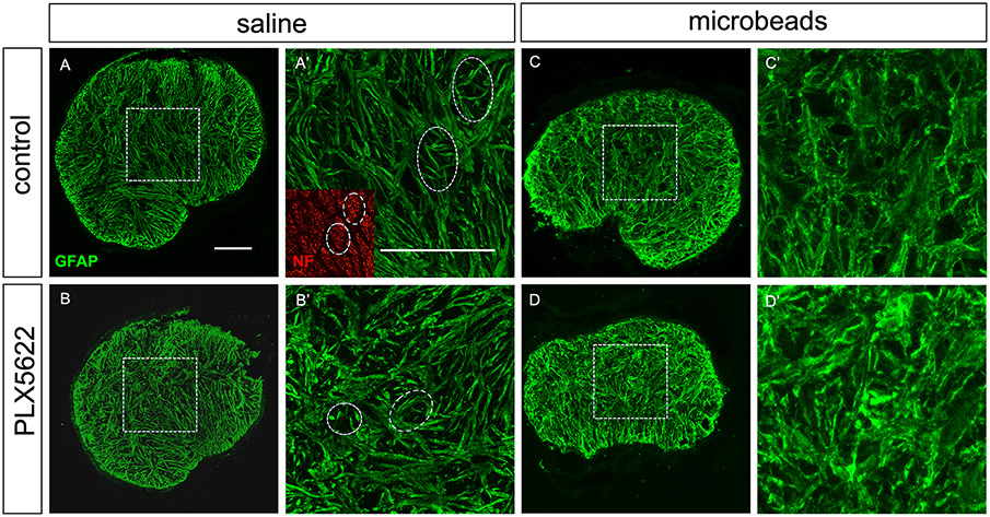
Astrocyte morphology. A-B, transverse sections of optic nerve heads (unmyelinated region) of saline-injected eyes stained with anti-GFAP antibodies. Microglia depletion by itself did not change astrocyte morphology. A’ and B’ show higher-magnification views of the boxed areas in A and B. Glial tubes through which ganglion cell axon bundles pass are indicated with white ovals. The inset in A’ shows axons stained with an antibody to neurofilament heavy chain (NF). C-D, astrocytes after microbead injection show the typical morphological signs of reactivity (process thickening and loss of glial tubes), but there was no difference between the control and the microglia-depleted groups. E, astrocyte process thickness was measured in optic nerves from 4 mice per group.

