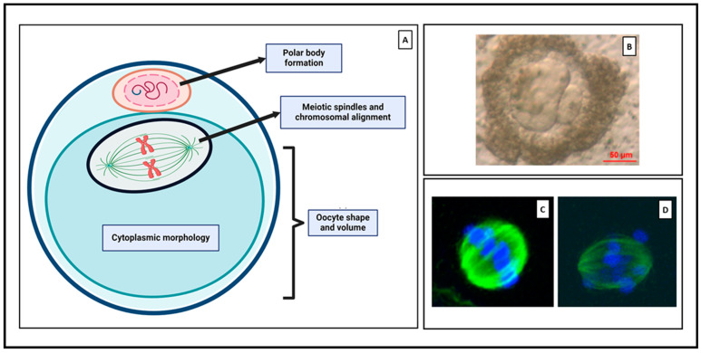
Figure 4.
(A) Schematic representation of morphological parameters used to assess oocyte competence. Created with BioRender.com. (B) Bright field image showing a MII oocyte with and enlarged abnormal polar body. Reproduced with permission from [10]. Confocal images displaying (C) equatorially aligned chromosomes (blue) and meiotic spindles (green), and (D) chromosomal misalignment. Reproduced with permission from [128].

