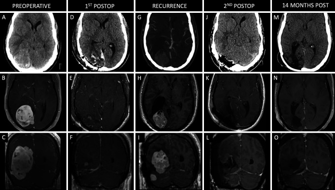
FIG. 2.
Case 2. Preoperative axial noncontrast CT (A) and axial (B) and coronal (C) contrast-enhanced T1-weighted MRI showing a large enhancing lesion centered along the right occipital convexity. Postoperative axial noncontrast CT (D) and axial (E) and coronal (F) contrast-enhanced T1-weighted MRI following initial right occipital craniotomy for GTR of the lesion. Axial contrast-enhanced CT angiogram (G) and axial (H) and coronal (I) contrast-enhanced T1-weighted MRI showing recurrence of the right occipital lobe lesion 76 months after the initial resection. Axial noncontrast CT (J) and axial (K) and coronal (L) contrast-enhanced T1-weighted MRI following right occipital/suboccipital craniotomy for GTR of the recurrent lesion. Axial noncontrast CT (M) and axial (N) and coronal (O) contrast-enhanced T1-weighted MRI scans 12 months after the second surgery (96 months after initial procedure).

