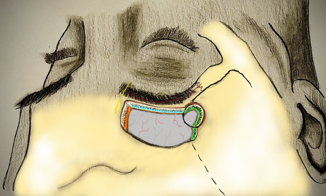
FIG. 2.

Illustration depicting the transciliary incision (red), supraorbital nerve (yellow), and the margins of the supraorbital ridge osteotomy (blue), medial edge of the osteotomy (orange), and sphenoid wing of the osteotomy (green). In addition to drilling of the orbital rim overhang, the modifications include drilling of the medial edge of the inner table to enhance the cross-court corridor for better visualization of the contralateral anterior cranial fossa in large anterior cranial base tumors. Additionally, drilling the inner table of the lateral limit of craniotomy facilitates sphenoid wing undercutting, which increases the surgical freedom to split the sylvian fissure and increase the exposure over the middle fossa in addition to the anterior skull base. Orbital rim can also be completely removed. However, in our case series, we did not see the need for complete removal of the orbital rim.
