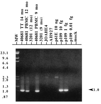
FIG. 2.
PCR analysis of cells from cat 40681. Nested PCR with a first-round primer pair specific for exogenous FeLV (FeLV-pol-5/FeLV-U3-2B) and a second-round primer pair specific for 81T variants (FeLV-81T-Ins/FeLV-U3-4) was performed on 100 ng of genomic DNA template (approximately 5 × 10⁴ cells) isolated from the following cat 40681 samples: thymic tumor (TT) harvested at necropsy (14 months [mo] p.i.) and PBMC from 12 and 9 months p.i. Additionally, 100 ng of genomic DNA from JOAHE4, the cell line used to generate the inoculum for cat 40681, was tested. Control templates included genomic DNA from the following sources: 3201 feline T cells that had been isolated in parallel with cat 40681 PBMC samples at 12 and 9 months p.i. (3201 [12 mo] and 3201 [9 mo], respectively), and uninfected AH927 feline fibroblasts. Plasmid control templates included 10 ng of p61E and a dilution series (10, 1, and 0.1 fg) of pEET(TE)-109 (p109). A reaction mixture containing water instead of DNA was included (mock). Second-round PCR product was electrophoresed through 1% agarose, stained with ethidium bromide, and photographed under UV light. Multiple amplifications using different primer pairs yielded similar results. Mobility of the molecular weight standards (MW), which are a mixture of phage lambda DNA cut with HindIII and phage φX174 DNA cut with HincII, are indicated with their corresponding sizes in kilobases on the left figure. On the right, the arrowhead indicates the position and size of the expected 1-kb amplimer.

