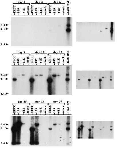
FIG. 5.
Southern blot analysis of proviral load and UVD during infection. 3201 T cells were infected with EECC, EET(TE)-109 (109), or 61E at an MOI of 0.005 or were mock infected, and genomic DNA was harvested at days 2, 4, 6, 8, 10, 12, 15, 19, and 37 p.i. In this experiment, which is similar to the one depicted in Fig. 3, cytopathicity was first apparent at approximately day 10. Twelve micrograms of DNA was digested with KpnI and analyzed by Southern blotting with probe exU3. Also included as a positive control was 4 μg of genomic DNA from the bone marrow (BM) of cat 1668, which died with immunosuppression and T-cell lymphoma after experimental infection with a mixture of 61E and 61B. This DNA sample was previously shown to harbor high levels of UVD in a similar analysis (28). The positions of the 3.4-kb fragments [corresponding to the internal KpnI fragment of 61E and EET(TE)-109] and 2.1-kb fragments (corresponding to the internal KpnI fragment of EECC) are indicated by arrowheads on the left. Also indicated with an arrowhead (labeled 0.4) is the position of the UVD fragments. The panels at the right indicate a longer exposure (days 2 to 6 p.i.) and shorter exposures (days 8 to 37 p.i.) of each Southern blot shown on the left.

