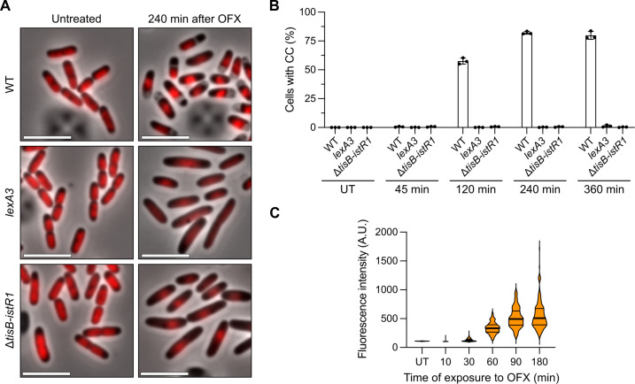
Fig. 2. The SOS-induced TisB toxin is responsible for cytoplasmic condensation during ofloxacin treatment.
Results are representative of three biological replicates. (A and B) WT, lexA3, and ΔtisB-istR1 cells encoding a hupA-mCherry fusion were grown in the microfluidic device for 60 min followed by perfusion with a medium containing OFX for 360 min. Image acquisition was performed every 15 min. (A) Representative images of the time-lapse experiment before and 240 min after the addition of OFX. Scale bars, 5 μm. (B) Percentage of cells in which CC was detected after addition of OFX. Between 297 and 1121 cells were analyzed for each sample. The data points are means ± SD. (C) Induction of transcriptional tisB reporter during OFX treatment. WT cells encoding a ptisB-gfp fusion were treated with OFX. At indicated times, cells were spotted on pads and imaged. Between 193 and 975 cells were analyzed for each sample. The data points are median (solid line) ± quartiles (dashed line).

