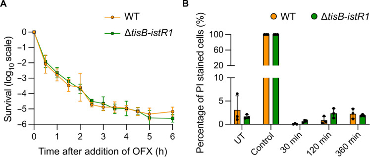
Fig. 6. Cytoplasmic condensation is not implicated in OFX-mediated lethality.
Cells were grown to an OD600nm ~ 0.3 and treated with OFX. Results are representative of three biological replicates. Data points are means ± SD. (A) Time-kill curve of WT and ΔtisB-istR1 strains. Culture samples were diluted and plated on LB agar plates. (B) Quantification of propidium iodide (PI)–stained WT and ΔtisB-istR1 cells. At indicated times, culture samples were incubated with PI for 15 min, spotted on agarose pads, and imaged. Isopropanol (70%) was used as positive control. Syto9 was used as a counterstain. Between 338 and 5830 cells were analyzed for each sample.

