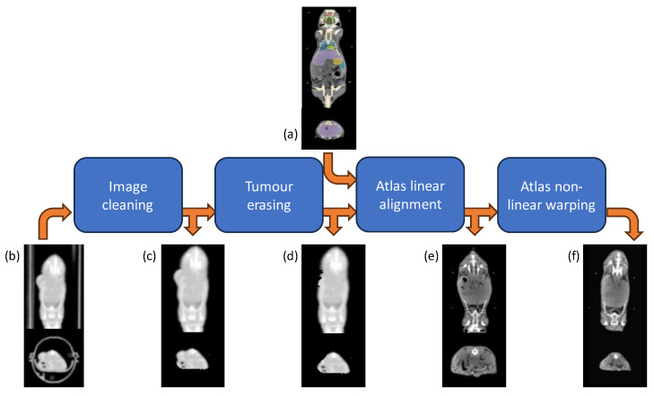
Figure 3.
Atlas-based segmentation workflow. Each original CT scan (b) underwent first pre-processing for animal holder removal (c) and tumor erasing (d); then the multi-organ segmentation was accomplished by warping the Digimouse CT atlas (a) through an affine transformation (e), followed by a B-spline and thin-plate spline mappings (f).

