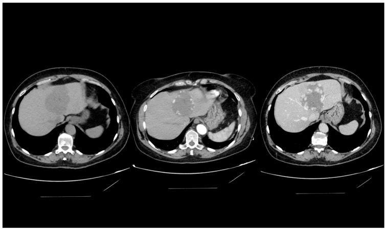
Figure 2.
Computed tomography imaging of a giant hepatic hemangioma. The sequence illustrates a precontrast image on the left, an arterial phase image in the center, and a venous phase image on the right. This series effectively demonstrates the delayed contrast filling from the tumor’s periphery, characteristic of a hepatic hemangioma.

