. 2024 Mar 27;12:RP91507. doi: 10.7554/eLife.91507

Figure 5. Hydrogen-deuterium exchange (HDX) assays survey conformation selection among compounds in the extracellular signal-regulated kinase-2 (ERK) inhibitor panel.

HDX measurements were performed with representative inhibitors shown in Figure 4, chosen for variations in their left-side, central scaffold, and right-side substituents. Time courses show deuterium uptake at the (A) DFG motif (peptide 161–168: LKICDFGL), (B) P+1 segment (peptide 191–198, YRAPEIML), and helix αF (peptide 203–210: YTKSIDIW). Enhanced HDX protection (strongly decreased uptake) in each segment by inhibitors #5, #6, #8, and #16 (blue) suggest properties of conformation selection for the R-state, while lower protection by inhibitors #4 and #15 (gray) suggest retention of conformational exchange. Inhibitor #1 (cyan) shows HDX properties intermediate to these two groups. HDX time courses for the full set of 17 inhibitors are shown in Figure 5—figure supplement 1 and Figure 5—figure supplement 2.

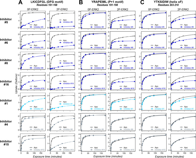
Figure 5.

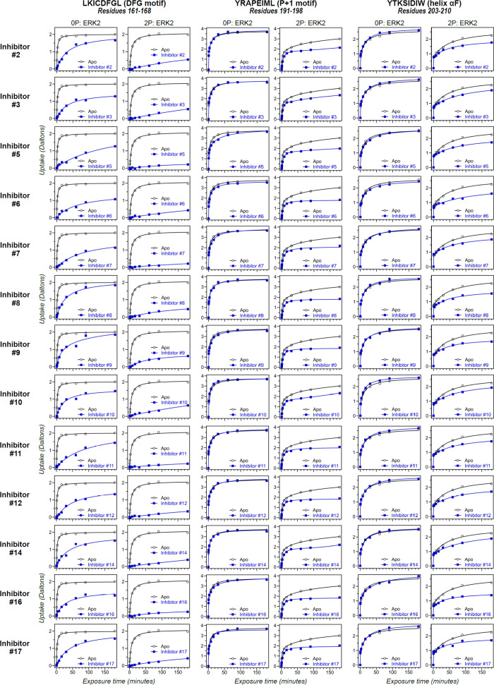
Figure 5—figure supplement 1. Differential hydrogen-deuterium exchange (HDX) responses to new inhibitors were surveyed in this study.

Figure 5—figure supplement 1.

Effects of binding the full set of inhibitors #1-#17 on deuterium uptake into the DFG motif (peptide 161–168: LKICDFGL), P+1 segment (peptide 191–198, YRAPEIML), and helix αF (peptide 203–210: YTKSIDIW), expanding the selected subset in Figure 5. Open symbols show deuterium uptake in 0P- or 2P-ERK2 apoenzymes. Closed symbols show deuterium uptake in 0P- or 2P-ERK2 complexed with inhibitors. Thirteen inhibitors (#2, #3, #5–12, #14, #16, and #17; blue) show HDX uptake patterns for 2P-ERK2 that are similar to Vertex-11e (VTX11e) and BVD523, suggesting conformation selection for the R-state.
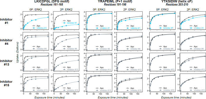
Figure 5—figure supplement 2. Differential hydrogen-deuterium exchange (HDX) responses to new inhibitors were surveyed in this study.

Figure 5—figure supplement 2.

Effects of binding the full set of inhibitors #1-#17 on deuterium uptake into the DFG motif (peptide 161–168: LKICDFGL), P+1 segment (peptide 191–198, YRAPEIML), and helix αF (peptide 203–210: YTKSIDIW), expanding the selected subset in Figure 5. Open symbols show deuterium uptake in 0P- or 2P-ERK2 apoenzymes. Closed symbols show deuterium uptake in 0P- or 2P-ERK2 complexed with inhibitors. Three inhibitors (#4, #13, #15; gray) show HDX patterns similar to GDC0994, consistent with R↔L exchange. One inhibitor (#1; cyan) shows significant HDX protection in the DFG motif, but only marginal protection in the P+1 segment or helix αF, suggesting behavior intermediate between Vertex-11e (VTX11e)/BVD523 and GDC0994.