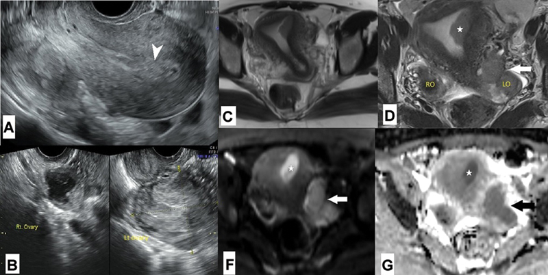
Fig. 6.

A 35-year-old who was being treated for primary infertility had ( A, B ) transvaginal ultrasound showing endometrial thickening (arrowhead) and US-O-RADS 4 left ovarian mass. Initial endometrial biopsy revealed endometrial hyperplasia. ( C, D ) Axial T2-weighted magnetic resonance imaging and ( E, F ) diffusion-weighted imaging with apparent diffusion coefficient map showed intermediate signal diffusion restricting eccentric soft tissue mass in the endometrial cavity near the left cornua of the uterus (*) with no myometrial invasion and an irregular lobulated predominantly solid diffusion restricting left ovarian mass (arrow) suggestive of malignant left ovarian mass. The right ovary was normal. There was no obvious peritoneal disease. The patient underwent total abdominal hysterectomy, bilateral salpingo-oophorectomy, omentectomy, and pelvic and para-aortic lymphadenectomy. Surgical histopathology revealed International Federation of Gynecology and Obstetrics (FIGO) 1A grade 1 endometroid endometrial carcinoma and FIGO 1C2 endometroid adenocarcinoma of the ovary. She was given adjuvant chemotherapy following the surgery.
