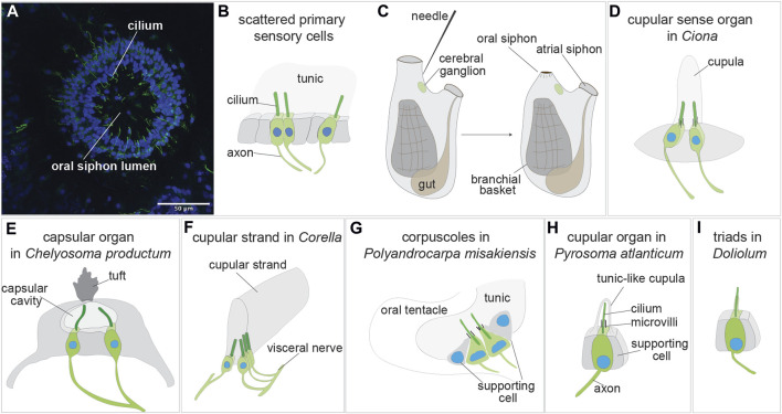
FIGURE 3.
Sensory organs based on primary sensory cells in adult tunicate (see Caicci et al., 2013; Manni and Pennati, 2015; Anselmi et al., 2022). (A) Confocal imaging of the primary sensory cells stained with anti-alpha tubulin (green) labelling nerves and Hoechst (blue) in the oral siphon of B. schlosseri. (B) Scattered primary sensory cells.) (C) Illustration of the primary sensory cell stimulation in the “siphon stimulation test”. (D) Cupular sense organ in Ciona. (E) Capsular organ in Cheliosoma productum. (F) Cupular strand in Corella. (G) Corpuscles in Polyandrocarpa misakiensis. (H) Cupular organ in Pyrosoma atlanticum. (I) Triads in Doliolum nationalis.

