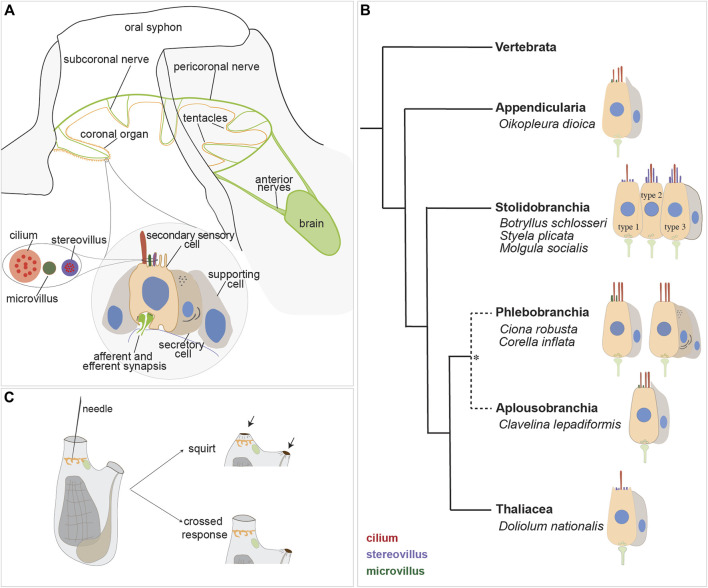
FIGURE 4.
Secondary sensory cells in the adult tunicates. (A) Location and main features of the coronal organ in tunicates. The organ is composed of a continuous row of cells on the oral tentacles and the velum (orange). Each sensory cell makes synapses with the subcoronal nerves (two per tentacle, close to the coronal organ) that are branches of the pericoronal nerve (green). The latter is a mixed nerve, connected to the brain through the anterior nerves. Sensory cells (pink) are flanked by supporting cells (grey); in some enterogona species, also secretory cells (violet) can be recognised. Stereovilli are apical, finger-like, long structures, composed of parallel actin filaments connected to the cell cytoskeleton; microvilli are thinner than stereovilli, with less abundant actin microfilaments. (B) Comparative schematic illustration showing the coronal organ variability in some representatives of tunicate groups. Stolidobranchia ascidians display the greatest complexity in the sensory apical bundle, which can be composed of microvilli or stereovilli, the latter also graded in length. * The monophyly of Phlebobranchia is disputed [see (DeBiasse et al., 2020)]. (C) Responses obtained after a strong (upper) and a light (bottom) stimulation of the coronal cells. The latter response is detected in the “tentacle stimulation test”.

