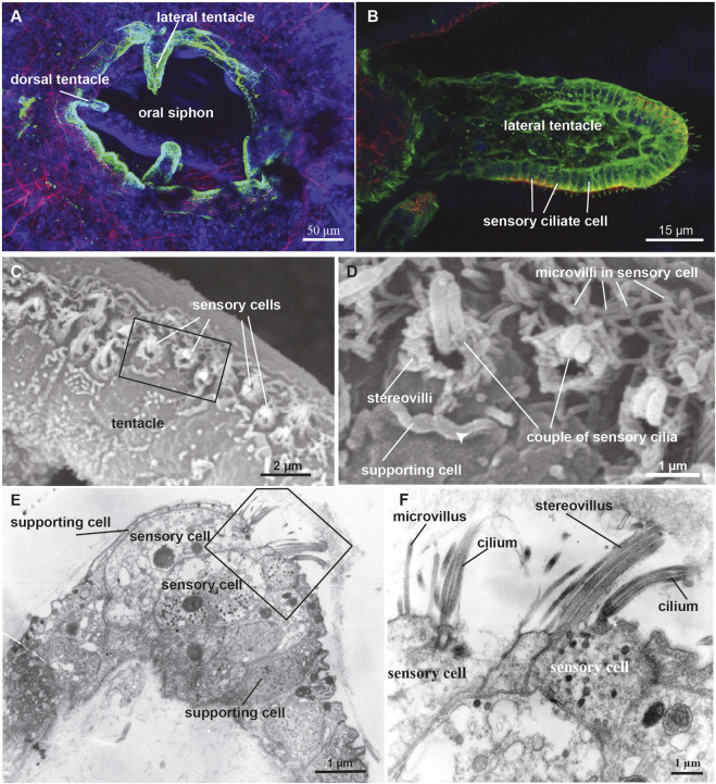
FIGURE 5.
(A,B) Confocal pictures of the B. schlosseri oral siphon and tentacles stained with anti-alpha tubulin (green) labelling nerves, phalloidin (red) labelling cytoplasmatic actin and dapi (blue) labelling cell nuclei. (C,D) Scanning electron microscopy showing the coronal organ of Molgula socialis. Squared area in C is enlarged in D. The organ is composed of a row of 1-2 sensory cells (recognisable by their hair bundle) flanked by supporting cells characterized by an apical cytoplasmic crista (arrowhead). Two types of sensory cells can be recognised: with a couple of cilia surrounded by graded stereovilli (type 3), and with a single cilium surrounded by microvilli (type 1). (E,F) Transmission electron microscopy showing a transverse section of the coronal organ of M. socialis. Squared area in E is enlarged in F to show the different apical bundle structure: two sensory cells at left display microvilli (type 1), whereas the sensory cell at right possesses stereovilli (type 2 or 3).

