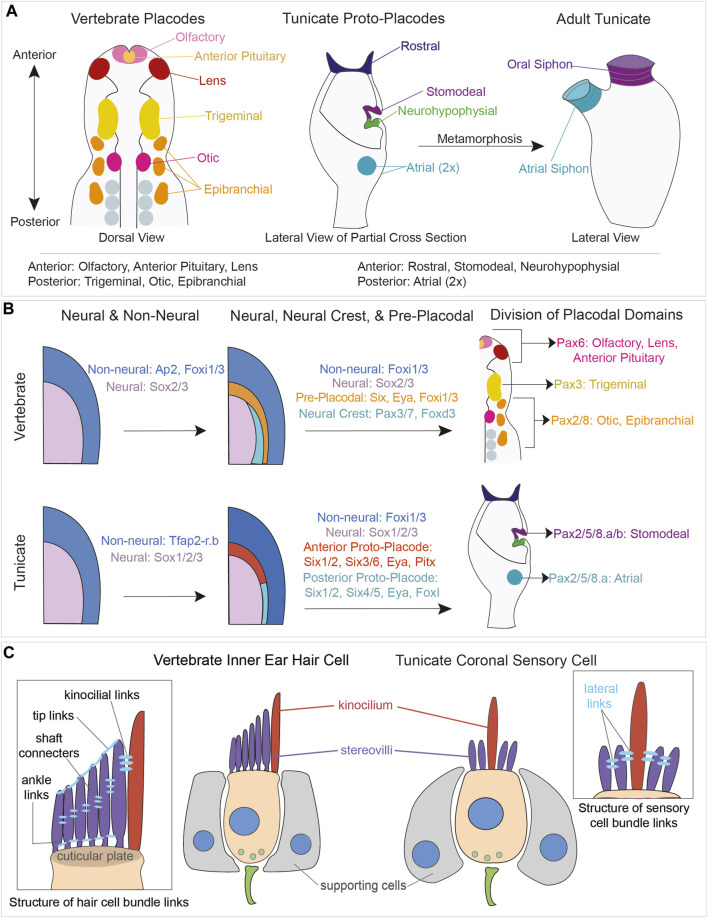
FIGURE 6.
Comparison of vertebrate placodal and tunicate proto-placodal development and vertebrate hair cell and tunicate coronal sensory cell structures. (A) Schematic of vertebrate placodes compared to tunicate proto-placodes. The anterior placodes include the olfactory, anterior pituitary, and lens placodes. The posterior placodes include the trigeminal, epibranchial, and otic placodes. Tunicates have three anterior proto-placodes:, the rostral, stomodeal, and neurohypophysial placodes. Tunicates have two posterior atrial proto-placodes. Following metamorphosis, the stomodeal proto-placode will give rise to the oral siphon and the atrial proto-placodes will fuse to form the atrial siphon. (B) Conservation of genes expressed during vertebrate placodal and tunicate proto-placodal development. Several key genes involved in placode development appear to be conserved. (C) Comparison of hair cells from vertebrates and coronal sensory cells from tunicates. Vertebrate hHair cells (tan) are flanked by supporting cells (gray). Sensory cells possess kinocilium (red) and stereovili (purple) that are connected together by different links.

