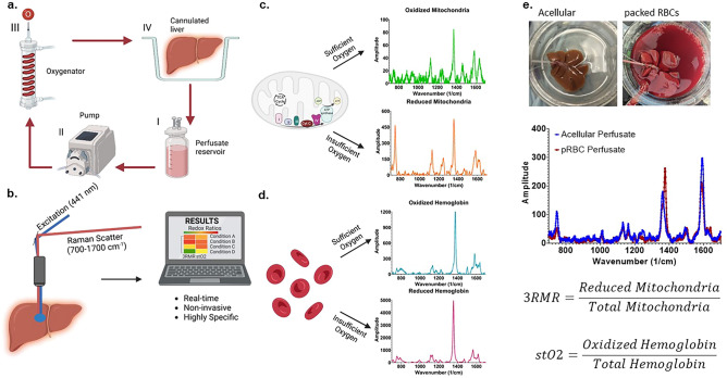
Figure 1.
Resonance Raman Spectroscopy predicts functional oxygenation of liver tissue during machine perfusion. (a) Schematic of machine perfusion of livers where a perfusate(I) with or without red blood cells as oxygen carriers is circulated by a pump (II) to an oxygenator (III). The oxygenator consists of a gas-permeable tubing that carries the perfusate surrounded by oxygen at a higher ambient partial pressure than atmosphere. This allows the perfusate to be oxygenated before it is supplied to a cannulated liver (IV). (b) The principle of our custom approach to measuring mitochondrial redox state in the tissue non-invasively using a resonance Raman spectroscopy device. A 441 nm excitation laser is used to excite molecules with a porphyrin ring (such as mitochondrial complexes, cytochromes, and hemoglobin) that produce a resonance Raman spectrum. (c) The spectrum of oxidized and reduced isolated mitochondria. (d) The spectrum of reduced and oxidized hemoglobin. (e) Two rat livers that are sitting in the perfusion bowl that are supplied with oxygen either with or without RBCs. Directly below the livers are the RRS spectrum from each liver. This spectrum is deconvoluted into its constituent molecular signatures using pre-recorded libraries as shown in c and d. The deconvolution coefficients for the reduced mitochondria is averaged with the sum of reduced and oxidized mitochondria to quantify the redox state of tissue and called the 3RMR value. Similarly, the oxygen saturation of hemoglobin in the tissue is also obtained by taking the ratio of oxidized hemoglobin coefficient with the total coefficients of oxidized and reduced hemoglobin.

