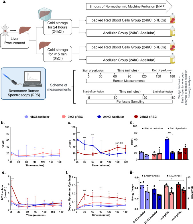
Figure 2.
3RMR with lactate, energy charge, and NAD:NADH ratio predict metabolic recovery dynamics during machine perfusion. (a) Schematic of the experiments and test groups. Each liver is either stored at 4 °C for 24 h or perfused immediately after recovery from the rat. It may then be machine perfused with either a packed RBC based perfusate or an acellular perfusate for 3 h at 37 °C. During this time the perfusate is sampled every 60 min for blood gas, blood chemistry, and ALT/AST analysis. Raman measurements are also taken from the surface of the livers every 30 min. At the end of the perfusion, wedge biopsies from the liver are either flash frozen for energetic tests or stored in formalin for histology. (b) Low 3RMR values throughout the 3 h of perfusion that are statistically indistinguishable between 0hCI acellular (n = 4) and 0hCI pRBC (n = 4) groups. (c) Significantly higher 3RMR value immediately after reperfusion of 24hCI acellular group (n = 3) compared to 24hCI pRBC group (n = 4). The 3RMR value decreases continuously during perfusion and becomes statistically indistinguishable around 90 min of perfusion. This trend reverses after 90 min indicating possible dysfunction of mitochondrial electron transport chain, however remaining statistically the same. (d) The comparison between 3RMR values at the start of perfusion and at the end of perfusion for each of the tested conditions. The p-values that are marked for each pair show statistically significant lower 3RMR values for acellular perfusate while those with packed RBCs are statistically the same. (e) Lactate levels during the 3 h of perfusion for all four conditions. That remain low and indistinguishable for all the tested conditions during perfusion. (f) Oxygen uptake rate for all four groups during perfusion. The oxygen uptake for 24hCI pRBC group is the highest which reflects the higher oxygen demand that is satisfied by the higher supply of oxygen bound to RBCs. OUR between the other three groups is comparable with only slightly higher values in the 0hCI pRBC group. (g) Energy charge and NAD:NADH values for all the four conditions tested. These were obtained from flash frozen wedge biopsies from the livers at the end of 3 h of perfusion. While not statistically significant, the ratios for machine perfusion of 24-h cold ischemic livers perfused with acellular perfusate were slightly lower than the other conditions, indicating potential slower recovery of oxidative phosphorylation. We plotted median ± IQR for all the variables. Statistical significance levels as follows *p < 0.05, **p < 0.01, ***p < 0.005.

