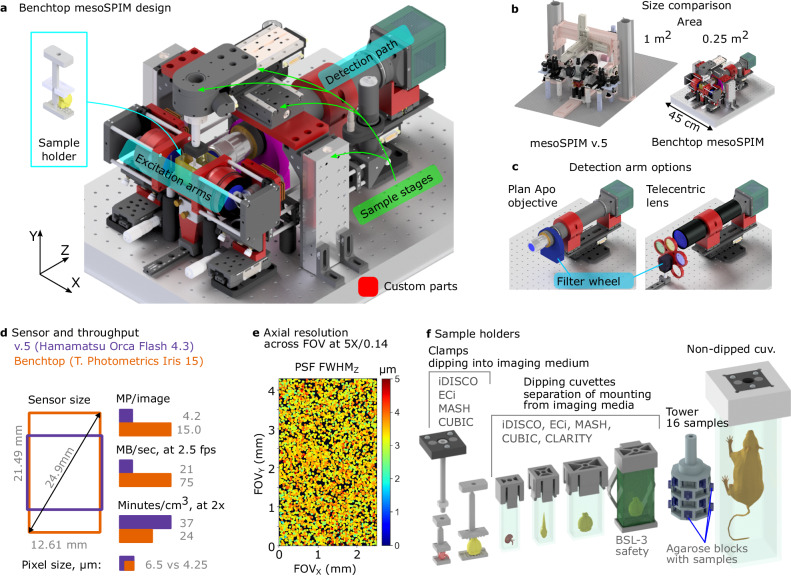
Fig. 1. The Benchtop mesoSPIM design and an application example.
a CAD model of the microscope with main modules labeled: excitation arms, detection path, sample stages, and sample holder. Modified or custom-made parts are red. b Size comparison between mesoSPIM v.5 and Benchtop systems. c Detection arm can be equipped with a plan apochromatic objective (2×–20× magnification, tube lens not shown) or a telecentric lens (0.9×–2×) depending on the application, with a corresponding filter wheel and set of filters. The detection arm is mounted on a focusing stage. d Comparison of sensor size, pixel size, pixel count per image, and imaging throughput between v.5 (Hamamatsu Orca Flash4.3 camera) and Benchtop (Teledyne Photometrics Iris 15). e The Benchtop mesoSPIM axial resolution in ASLM mode across the FOV, at magnification 5× and NA 0.14. The full width at half-maximum of the point-spread function along the z-axis (FWHMZ) is color-coded from 0 to 5 µm. The resolution was measured with 0.2 µm fluorescent beads embedded in agarose immersed high-index medium (RI = 1.52). f CAD models of custom 3D printed sample holders that accommodate samples from 3 mm to 75 mm.

