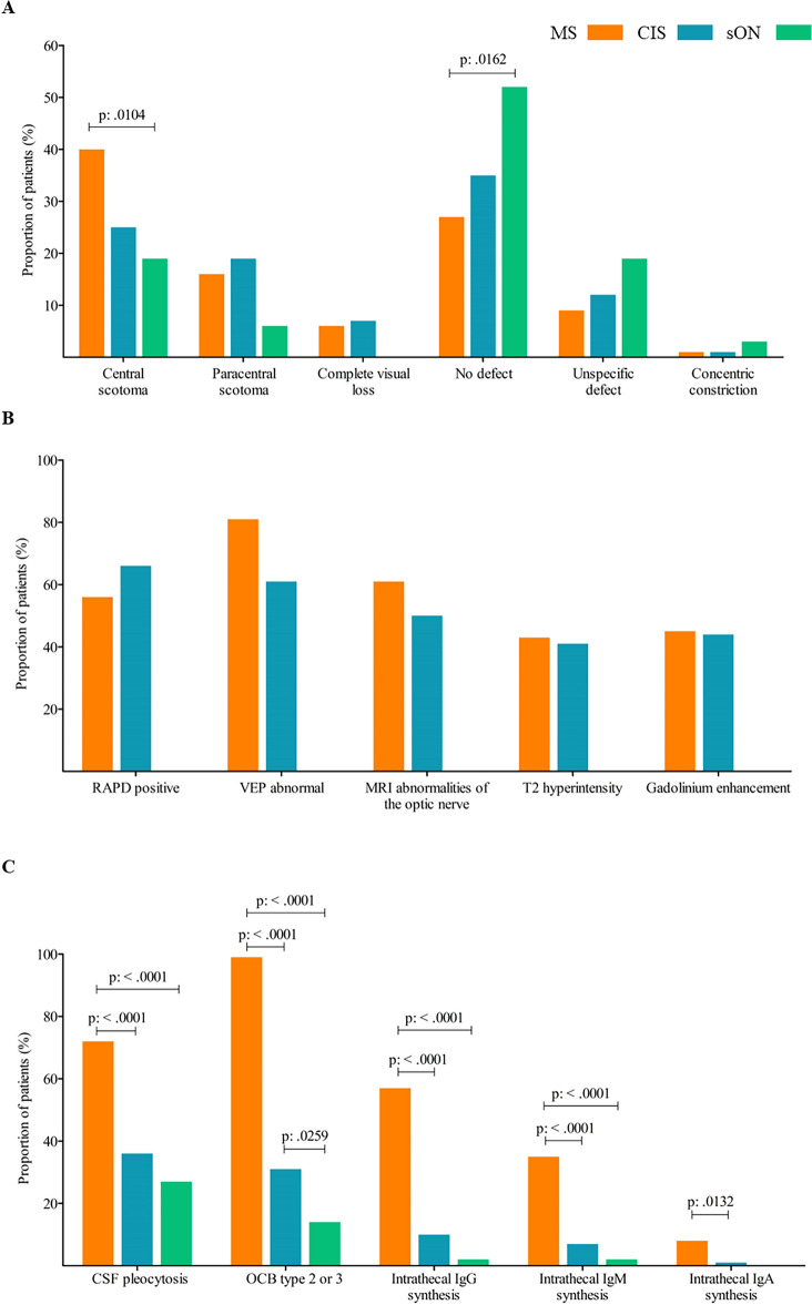
Figure 3.
(A) Percentage of patients with the respective perimetry abnormalities for each group. (MS—optic neuritis, CIS—optic neuritis, suspected optic neuritis). MS, multiple sclerosis; CIS, clinically isolated syndrome; sON, suspected optic neuritis. (B) Proportion of patients with objectifiable findings such as RAPD, abnormalities in MRI of the optic nerve or VEP for the groups MS—optic neuritis and CIS—optic neuritis. By definition, the sON group did not show any objectifiable abnormalities for optic neuritis, as this would at least have meant classification in the CIS group. RAPD, relative afferent pupillary deficit, VEP, visual evoked potential; MRI, magnetic resonance imaging. MS, multiple sclerosis; CIS, clinically isolated syndrome. (C) Percentage of patients with the respective CSF finding for each group. (MS—optic neuritis, CIS—optic neuritis, suspected optic neuritis). CSF, cerebrospinal fluid; OCB, oligoclonal bands; MS, multiple sclerosis; CIS, clinically isolated syndrome; sON, suspected optic neuritis.

