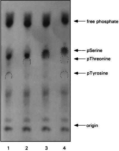
FIG. 2.
I3 is phosphorylated on serine residues. Cells were infected with wt virus (MOI of 15) and metabolically labeled with 32Pi. Lysates were prepared and subjected to immunoprecipitation with anti-I3 serum. Liberated immune complexes were subjected to trichloroacetic acid precipitation and then to hydrolysis with HCl. The hydrolysates were mixed with phosphoamino acid markers either individually (lanes 1 through 3) or as a mixture (lane 4). Samples were then applied to thin-layer cellulose plates and resolved by high-voltage electrophoresis. Markers were visualized by color development with ninhydrin; radiolabeled phosphoamino acids derived from I3 were visualized by autoradiography. The dotted wickets indicate the positions of migration of the phosphoserine (pSerine), phosphothreonine (pThreonine), and phosphotyrosine (pTyrosine) markers. The position of migration of free phosphate is indicated, as is the application origin.

