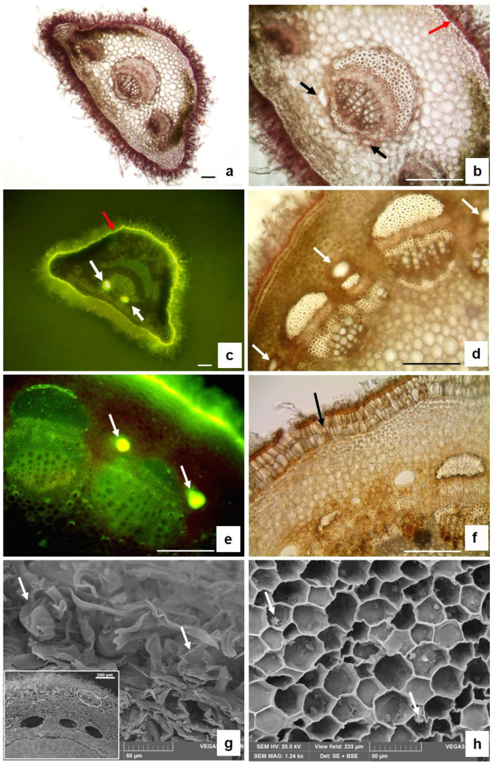
Figure 4.
LM (a–f) and SEM (g,h) micrographs of transversal sections of a leaf petiole (a–c) and young stem (d,h). (a) General petiole anatomy; (b) detail of the petiole showing two secretory ducts on both sides of the main vascular bundle (black arrows) and a thick cuticle over the epidermis, stained red with Sudan III (red arrows); (c) brilliant yellow fluorescence of the lipophilic substances of the cuticle (red arrows) and of the essential oil inside the secretory ducts stained with FY (white arrows); (d) young stem anatomy revealing the presence of secretory ducts in the cortex near the vascular bundle (white arrows); (e) brilliant yellow fluorescence of the essential oil inside secretory ducts stained with FY (arrows); (f) periderm (black arrow) in the older stem; (g) glandular trichomes on the stem surfaces (white arrows); the insert shows the location of the glandular trichomes; and (h) small crystal druses inside the pith parenchyma cells (white arrows).

