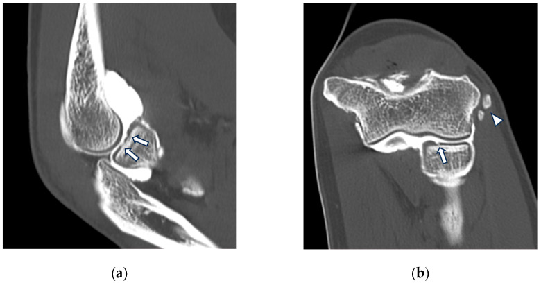
Figure 3.
CT arthrography of a patient with chronic elbow instability. (a) Sagittal image shows pitting and fissuring of radial head cartilage, involving >50% of its thickness (grade III) (white arrows); (b) coronal reformat of the same patient shows a focal full-thickness cartilage defect of the anteromedial radial head (grade IV) (white arrow). In the same image, mature calcifications can be seen at the insertion of the common extensor tendon (white arrowhead).

