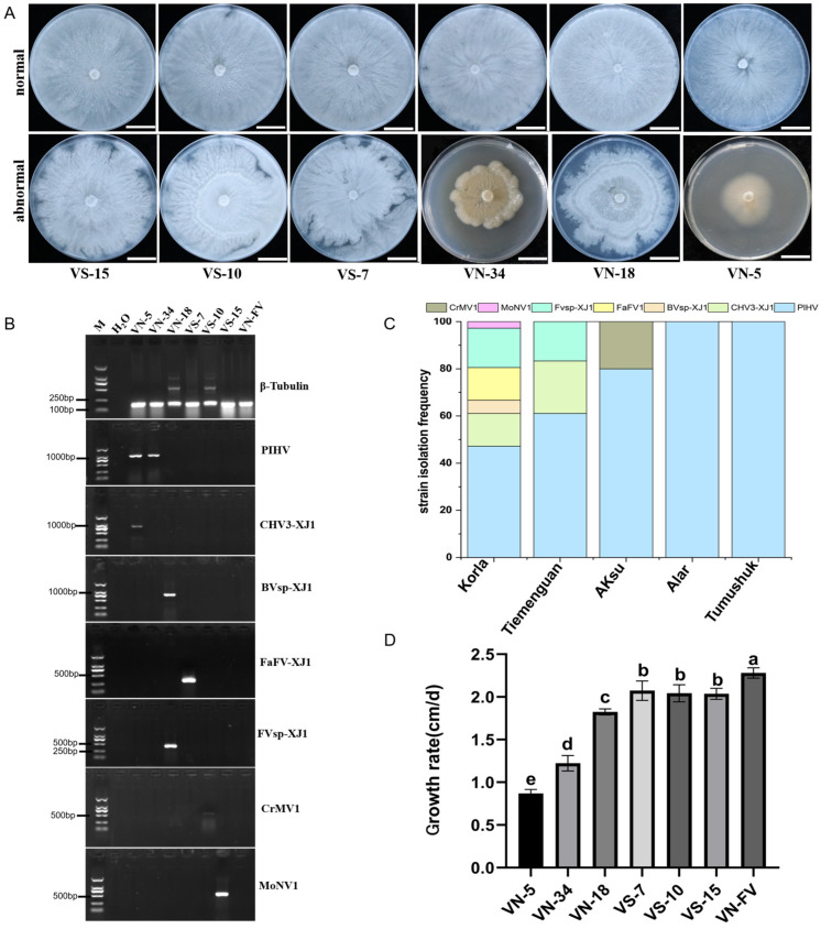
Figure 2.
Detection of the seven mycoviruses and their effect on the growth of Valsa spp. (A) Colony morphology of Valsa spp. strains grown on PDA plates for 5 d (bars = 2 cm); (B) the detection of mycovirus contigs in different Valsa spp. strains. The internal reference gene β-Tubulin from Valsa spp. was used as the positive control, and ddH2O was used as the negative control. (C) Frequency of mycoviruses identified in different regions; (D) the growth rates of Valsa spp. strains harboring mycoviruses. The data are presented as means ± SD (n = 4). The different letters indicate a significant difference at p < 0.01 (determined via one-way ANOVA).

